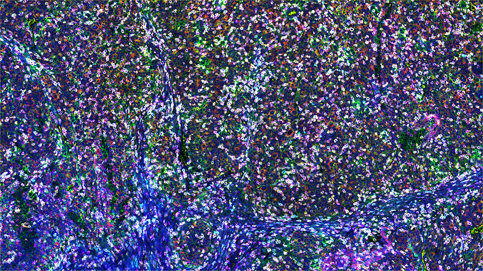
Figure legend: SignalStar multiplex immunohistochemical analysis of paraffin-embedded human gastric adenocarcinoma using Pan-Keratin (C11) & C0-0003-488 SignalStar Oligo-Antibody Pair #63566 (red), PD-L1 (E1L3N) & C0-0005-488 SignalStar Oligo-Antibody Pair #85646 (green), PD-1 (Intracellular Domain) (D4W2J) & C0-0008-594 SignalStar Oligo-Antibody Pair #35347 (yellow), Granzyme B (D6E9W) & C0-0009-594 SignalStar Oligo-Antibody Pair #15194 (orange), TIGIT (E5Y1W) & C0-0002-647 SignalStar Oligo-Antibody Pair #18288 (cyan), CD20 (E7B7T) & C0-0011-647 SignalStar Oligo-Antibody Pair #36775 (pink), CD3ε (D7A6E) & C0-0001-750 SignalStar Oligo-Antibody Pair #51754 (magenta), CD8α (D8A8Y) & C0-0004-750 SignalStar Oligo-Antibody Pair #62750 (white), and ProLong Gold Antifade Reagent with DAPI #8961 (blue). All fluorophores have been assigned a pseudocolor, as indicated. Staining was performed on the BOND RX autostainer by Leica Biosystems.
SignalStar Multiplex Immunohistochemistry (mIHC) is a tool for studying FFPE tissue samples, using antibodies, oligonucleotides, and fluorophores to identify which cells are present, where they are located, and what functions they perform.
SignalStar technology enables the detection of multiple phenotypic and functional targets while preserving spatial context and tissue architecture. These insights are critical for understanding how cells organize and interact to influence the tissue microenvironment that ultimately drive disease progression and response to therapy.
The resources below can provide support for using SignalStar technology in your research.